
Journey
One-way simulation from start to destination. The province/state and country narrow the search so the correct place is found even when several towns share a name. Only enter an average speed if you choose the custom option; by default the route engine estimates driving time itself. Route and elevation data: OSRM demo server, Nominatim geocoding and Open-Meteo elevation (Copernicus DEM 90 m). All free public services; occasional slow responses are normal.
Clinical Parameters
Axial length refines globe volume (scaled as (AL/24)³ of 7211 µL), which converts the fill percentage into an absolute bubble volume. Leave blank to use the standard globe volume. Injected gas concentration is not needed here: by travel time the bubble has equilibrated and obeys Boyle's Law regardless of its original composition. Concentration matters only for estimating the current fill, below.
Estimate current fill from surgical details
Bubble size projection. This estimator lets the surgeon project the bubble size on any post-operative day from the injected mixture, so travel can be planned for the day the fill drops into a safe range. Enter the concentration actually used, for example 22% SF₆, and the fill achieved at the end of surgery.
Non-expansile concentrations (about 20% SF₆, 16% C₂F₆, 12% C₃F₈) hold their injected volume; pure gas expands to its empirical maximum (SF₆ ×2, C₂F₆ ×3.3, C₃F₈ ×4). Intermediate concentrations are interpolated between those two anchors; concentrations at or below the non-expansile point are treated as non-expansile, with dissolution starting immediately. First-order estimates only: confirm the fill clinically where the result matters.
Advanced physiological constants
Why Altitude Matters After Gas Tamponade
Pars plana vitrectomy and pneumatic retinopexy frequently rely on an intraocular gas bubble to tamponade the retina against the retinal pigment epithelium while retinopexy matures. The healthy eye is filled with incompressible fluid and is essentially indifferent to barometric pressure. A gas-filled eye is not. A trapped bubble obeys Boyle's Law: as ambient pressure falls with ascent, the bubble expands, and because the scleral shell has limited compliance, that expansion is converted into a rise in intraocular pressure. Sustained IOP above 30 mmHg compromises ocular perfusion and risks retinal ischaemia, central retinal artery occlusion and pressure-induced optic neuropathy. Descent does the opposite: bubble compression produces hypotony, with its own risks of choroidal effusion and re-detachment.
For patients travelling home from Mbombela through the Mpumalanga escarpment, the practical question is not whether altitude changes matter, but by how much, for how long, and on which specific route. This calculator answers that question by simulating the eye minute by minute along the actual road profile.
The Physics Engine
1. Barometric pressure from elevation
Every sampled point on the route is converted from elevation to absolute atmospheric pressure using the standard barometric formula for the troposphere (sea-level pressure 760 mmHg, lapse rate 0.0065 K/m, standard temperature 288.15 K).
2. Boyle's Law for the bubble
Body temperature is constant, so the bubble behaves isothermally. Its volume at any moment is set by the absolute intraocular pressure, which is the ambient pressure plus the gauge IOP a tonometer would read.
3. Friedenwald ocular rigidity
The pressure-volume behaviour of the globe is exponential, not linear. Friedenwald showed in 1937 that the logarithm of IOP changes in proportion to volume change, scaled by the coefficient of ocular rigidity K. Surgery changes K substantially. Applying the ideal gas law to Friedenwald's equation, Simone and Whitacre derived effective rigidity coefficients of 0.0021 µL⁻¹ for vitrectomised eyes with cryopexy and 0.0013 µL⁻¹ for eyes with an encircling buckle, against roughly 0.0126 µL⁻¹ for the unoperated eye. Lower K means a more compliant globe that absorbs bubble expansion with a smaller pressure rise, which is why surgical history is a required input.
4. Goldmann aqueous dynamics: the eye's shock absorber
The eye is not a sealed vessel. The ciliary body secretes aqueous at about 2.5 µL/min and the trabecular meshwork drains it in proportion to the gradient between IOP and episcleral venous pressure (facility about 0.25 µL/min/mmHg). When the bubble expands and IOP rises, outflow accelerates and fluid leaves the eye, partially offsetting the expansion. This is why speed of ascent matters: a slow climb gives the meshwork time to decompress the eye, a fast climb does not. The simulator integrates this outflow at every time step.
5. The coupled solver
At each 15-second step the engine must reconcile three competing demands: the bubble wants to expand, the aqueous compartment has drained or accumulated a known volume, and the sclera converts the net volume change into pressure. Because IOP appears on both sides of the combined equation, it is solved numerically (bisection on the monotonic residual) at every step, using the exact exponential Friedenwald relation rather than a linearised approximation. Importantly, the simulation does not stop when the car does. If the eye arrives at the destination with pressure above 30 mmHg, the engine keeps integrating with the patient stationary at destination altitude until the pressure drops back below the 30 mmHg threshold, because the bubble remains expanded at that altitude and the eye can only decompress at the trabecular outflow rate. A patient who crests a pass five minutes before arriving does not stop accumulating ischaemic exposure at the front door; the time taken to fall below 30 mmHg after arrival is therefore added to the trip's total time above threshold. The same applies in reverse: an eye arriving in hypotony is followed until it refills out of the hypotonic range, and that recovery period counts toward the hypotony total.
6. The hypotonic feedback loop on descent
The least intuitive and most clinically important behaviour the model captures involves routes that descend before they climb. On descent the bubble compresses, IOP falls, and once it drops below episcleral venous pressure the trabecular meshwork stops draining entirely, acting as a one-way valve. The ciliary body keeps secreting, so the eye quietly overfills with aqueous. When the patient later climbs back out of the valley, the bubble re-expands into an eye that is now volumetrically overloaded, and the resulting spike can exceed anything predicted from the maximum elevation alone. This is why the calculator processes the full sequential topography of the route, and why a road that dips through a valley before climbing deserves as much scrutiny as the summit itself.
Gas Pharmacokinetics
Pure perfluorocarbon gases expand because of Dalton's law of partial pressures and Fick's laws of diffusion. A freshly injected pure bubble contains no nitrogen, oxygen or carbon dioxide, so steep concentration gradients drive those gases from the surrounding tissue and bloodstream into the bubble far faster than the large, heavy fluorinated molecules can diffuse out. The bubble grows until the partial pressures approach equilibrium, then dissolves with first-order kinetics as the tamponade gas slowly leaves. Wong and Thompson showed the disappearance phase plots as a straight line on a logarithmic scale, which is what the fill estimator on the calculator tab exploits.
| Gas | Peak expansion | Max expansion ratio (pure) | Non-expansile concentration | Approx. half-life | Intraocular duration |
|---|---|---|---|---|---|
| Filtered air | Immediate | 1× (non-expansile) | n/a | 1.3 days | 5 to 7 days |
| SF₆ | 24 to 48 h | 2× | ~18 to 20% | 4 to 6 days | 10 to 21 days |
| C₂F₆ | 36 to 60 h | 3.3× | ~16% | ~10 days | 28 to 35 days |
| C₃F₈ | 72 to 96 h | 4× | ~12% | ~35 days | 55 to 65 days |
Concentration and the non-expansile point
Whether a bubble expands at all depends on the injected concentration. Diluting the tamponade gas with air pre-loads the bubble with nitrogen and oxygen, flattening the diffusion gradients that drive expansion. At a specific concentration for each gas the inward and outward fluxes balance and the bubble holds its injected volume: Peters and colleagues measured this non-expansile, equilibrated concentration at approximately 12% for C₃F₈, and comparable work places SF₆ near 18 to 20% and C₂F₆ near 16%. This is why a surgeon performing a complete fluid-gas exchange uses a dilute mixture, while a pneumatic retinopexy uses a small volume of pure gas and relies on expansion. Crittenden and colleagues set out the principles for predicting expansion of intermediate mixtures; the calculator's estimator interpolates linearly between the non-expansile point and the empirical pure-gas maximum, which is adequate for planning purposes.
Two points deserve emphasis. First, injected concentration governs the expansion phase only: once the bubble has equilibrated with dissolved blood gases, a bubble that began as 22% SF₆ and one that began as pure SF₆ obey Boyle's Law identically, which is why the route simulation needs only the current fill. Second, dissolution is faster in vitrectomised eyes than in eyes with intact vitreous, so published half-lives are a guide rather than a guarantee, and the projected fill should be confirmed clinically before any consequential travel decision.
Clinical Thresholds and Air Travel Evidence
Why 30 mmHg, and why duration matters
Ocular perfusion depends on the difference between arterial pressure and IOP. As IOP climbs toward the perfusion pressure of the central retinal artery, blood flow to the inner retina and optic nerve head falls; sustained pressures above roughly 30 mmHg meaningfully erode perfusion reserve, and spikes into the 60 to 80 mmHg range can approach retinal artery closure outright. A two-minute excursion to 32 mmHg and forty minutes above 45 mmHg are very different clinical events, which is why the calculator reports both the peak and the cumulative minutes above threshold rather than a single number. On the other side, sustained hypotony below about 5 mmHg risks choroidal effusion, hypotony maculopathy and mechanical failure of the fresh retinopexy.
What the aviation studies showed
The most controlled human data come from simulated flight. Mills and colleagues placed nine post-vitrectomy patients in a hypobaric chamber replicating a commercial cabin altitude of about 7400 feet: eyes carrying only 10 to 15% residual gas showed a mean IOP rise of 109% during ascent, and after return to ground level the same eyes dropped to well below their baseline pressure, demonstrating both limbs of the pressure swing in a single session. Earlier animal work by Dieckert and colleagues found dangerous pressure rises with gas volumes as small as 0.25 ml, with transient central retinal artery occlusion observed, and Lincoff's paired 1989 papers characterised the compensation mechanisms and their limits. Kokame and Ing documented a tolerated low-altitude flight with a large bubble, underlining that outcome depends on the interaction of bubble size, altitude profile and time, not on any single factor. Road travel through the escarpment reaches cabin-equivalent altitudes, but with one decisive advantage the aircraft cannot offer: the driver can slow down, stop, or turn back, which is precisely the decision this tool is designed to inform.
How the flight mode works
The flight mode is included for academic interest only. Flying with an intraocular gas bubble is contraindicated, and no output of this simulation changes that: a patient should never fly with an intraocular gas bubble. The mode exists to demonstrate quantitatively why the contraindication holds across cabin altitudes, fill volumes and flight durations.
The calculator's flight mode applies the same physics engine to a pressure-equivalent altitude profile instead of a road elevation profile. The user enters the departure and arrival locations, and the ground elevation of each is retrieved automatically. The decisive quantity is the highest pressure-equivalent altitude reached at any point in the journey, not the destination: a patient starting at 900 m, flying with an 8 000 ft (2 440 m) cabin, and landing at 1 500 m experiences maximum bubble expansion at cruise, while the arrival ground elevation determines how much of that expansion persists after landing. Modern airliners typically hold cabin altitude near 6 000 ft; older or conservatively modelled cabins near 8 000 ft, the regulatory ceiling; a custom cabin altitude can be entered, and for non-pressurised aircraft the actual maximum flight altitude applies directly. The profile is modelled as a cabin climb over about 25 minutes, a cruise, and a descent over about 30 minutes ending at the arrival ground elevation, followed by the same post-landing settling phase used for road journeys.
Flight duration is a genuine variable, not a formality. On a short flight the eye carries most of its pressure spike through the whole cruise. On a long flight the aqueous outflow has hours to work at cruise cabin altitude, so the pressure partially equalises in the air; the exposure above 30 mmHg is concentrated in the first part of the cruise. The trade-off is that an equalised eye then meets the cabin descent with a bubble compressing below its equilibrium, which can convert the landing into a period of hypotony. Both limbs of this behaviour were observed directly in the hypobaric chamber study of Mills and colleagues, and both appear in the simulated pressure trace.
Model Assumptions and Limitations
The simulation assumes isothermal bubble behaviour, a fixed quantity of gas over the journey (no meaningful expansion or dissolution during the drive itself), linear inflow and outflow within physiological limits, a spherical single bubble, and standard-atmosphere barometric conditions. Injected gas concentration is deliberately not an engine input: once the bubble has equilibrated with dissolved blood gases, it obeys Boyle's Law regardless of its original composition, so concentration only informs the fill estimator. When axial length is supplied, globe volume is scaled as the cube of AL relative to a 24 mm reference eye, a geometric approximation. The model does not account for weather-related pressure variation, pseudofacility, choroidal volume shifts, or individual variation in outflow facility, which is substantially reduced in many post-surgical and glaucomatous eyes. Elevation data carries the resolution limits of the underlying digital elevation model (about 90 m horizontal), and road tunnels or cuttings may not be fully reflected.
Validation status. Every component of this model is drawn from published, peer-reviewed research: Friedenwald's pressure-volume relationship, the surgically altered rigidity coefficients of Simone and Whitacre, Goldmann aqueous dynamics, established gas pharmacokinetics and the standard barometric formula. The integrated route simulation, however, has not itself been validated against measured intraocular pressures in travelling patients. Its outputs are estimates for clinical decision support: a draft aid for the treating ophthalmologist, never a clearance to travel, and never the sole basis for a safety decision. They must be interpreted alongside examination findings, the actual measured IOP, gas fill assessment and the individual patient's optic nerve status.
References
- Friedenwald JS. Contribution to the theory and practice of tonometry. Am J Ophthalmol. 1937;20:985-1024.
- Simone JN, Whitacre MM. The effect of intraocular gas and fluid volumes on intraocular pressure. Ophthalmology. 1990;97(2):238-243.
- Thompson JT. Kinetics of intraocular gases: disappearance of air, sulfur hexafluoride, and perfluoropropane after pars plana vitrectomy. Arch Ophthalmol. 1989;107(5):687-691.
- Fromow-Guerra J, Sonda-Chávez GA, et al. The effect of altitude on intraocular pressure in vitrectomized eyes with sulfur hexafluoride tamponade by the Friedenwald method: rabbit animal model. BioMed Res Int. 2016;2016:7326160. PMID 27957500.
- Lincoff H, Weinberger D, Reppucci V, Lincoff A. Air travel with intraocular gas. I. The mechanisms for compensation. Arch Ophthalmol. 1989;107(6):902-906.
- Lincoff H, Weinberger D, Stergiu P. Air travel with intraocular gas. II. Clinical considerations. Arch Ophthalmol. 1989;107(6):907-910.
- Dieckert JP, O'Connor PS, Schacklett DE, et al. Air travel and intraocular gas. Ophthalmology. 1986;93(5):642-645.
- Kokame GT, Ing MR. Intraocular gas and low-altitude air flight. Retina. 1994;14(4):356-358.
- Dastiridou AI, Ginis HS, de Brouwere D, Tsilimbaris MK, Pallikaris IG. Ocular rigidity, ocular pulse amplitude, and pulsatile ocular blood flow: the effect of intraocular pressure. Invest Ophthalmol Vis Sci. 2009;50(12):5718-5722.
- Brubaker RF. Goldmann's equation and clinical measures of aqueous dynamics. Exp Eye Res. 2004;78(3):633-637.
- Altitude-associated intraocular pressure changes in a gas-filled eye. Retin Cases Brief Rep. DOI 10.1097/ICB.0000000000000852.
- Peters MA, Abrams GW, Hamilton LH, Burke JM, Schrieber TM. The nonexpansile, equilibrated concentration of perfluoropropane gas in the eye. Am J Ophthalmol. 1985;100(6):831-839.
- Wong RF, Thompson JT. Prediction of the kinetics of disappearance of sulfur hexafluoride and perfluoropropane intraocular gas bubbles. Ophthalmology. 1988;95(5):609-613.
- Mills MD, Devenyi RG, Lam WC, Berger AR, Beijer D, Lam SR. An assessment of intraocular pressure rise in patients with gas-filled eyes during simulated air flight. Ophthalmology. 2001;108(1):40-44.
- Abrams GW, Edelhauser HF, Aaberg TM, Hamilton LH. Dynamics of intravitreal sulfur hexafluoride gas. Invest Ophthalmol. 1974;13(11):863-868.
- Crittenden JJ, de Juan E Jr, Tiedeman J. Expansion of long-acting gas bubbles for intraocular use: principles and practice. Arch Ophthalmol. 1985;103(6):831-834.
- Jacobs P, Twomey J, Leaver P. Behaviour of intraocular gases. Eye. 1988;2:660-663.
- Killey FP, Edelhauser HF, Aaberg TM. Intraocular sulfur hexafluoride and octofluorocyclobutane: effects on intraocular pressure and vitreous volume. Arch Ophthalmol. 1978;96(3):511-515.
Routing © OSRM contributors. Geocoding and map data © OpenStreetMap contributors (ODbL). Elevation data © Open-Meteo / Copernicus DEM GLO-90.
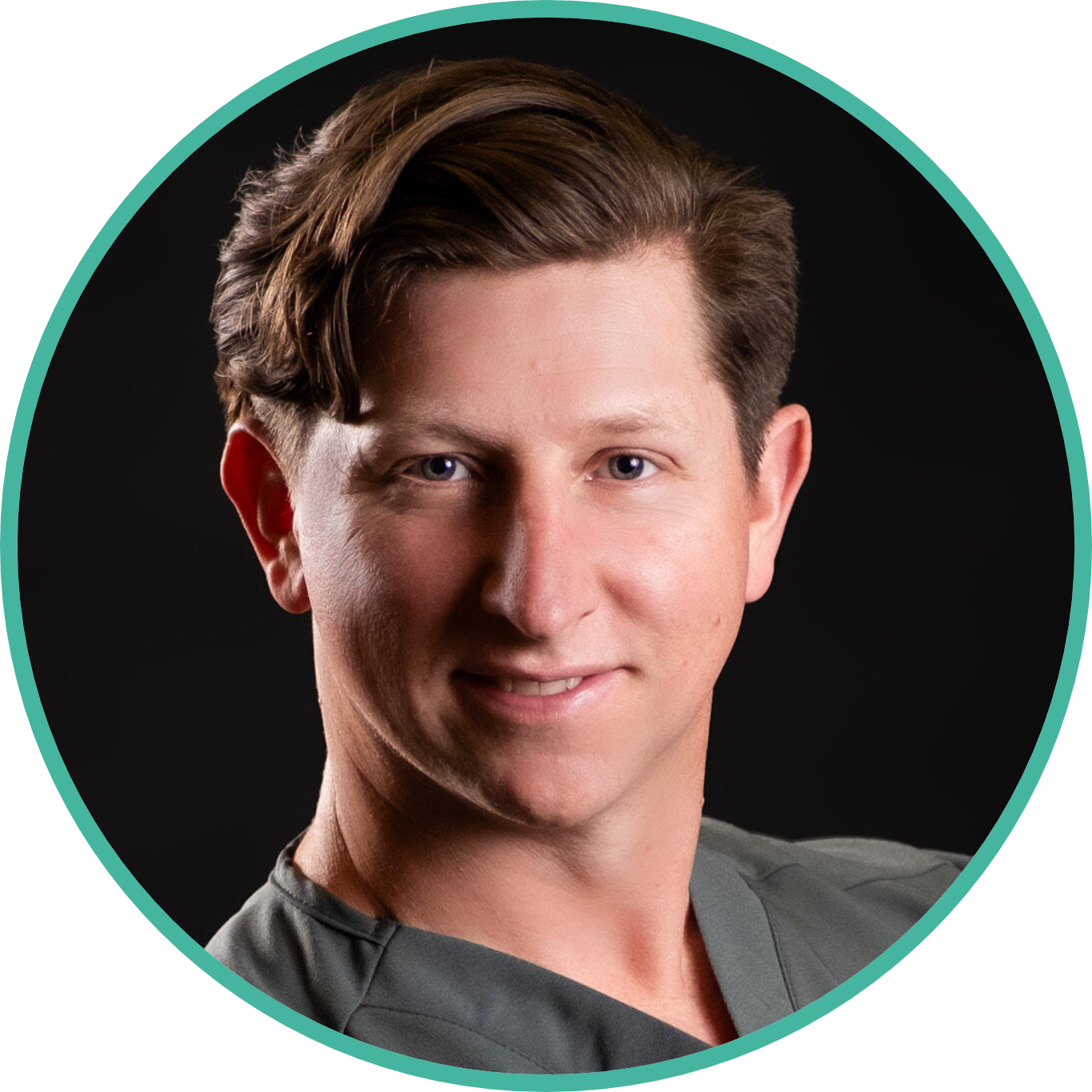
Dr Dean van der Westhuizen
Specialist Ophthalmologist and Vitreoretinal SurgeonPassionate about combining clinical ophthalmology with technology to reduce friction in patient care. This calculator was built to give vitreoretinal surgeons a route-specific, physics-based way to counsel patients with intraocular gas about travel over the escarpment, replacing blanket altitude rules with a simulation of the actual journey. It is shared freely with the vitreoretinal community; feedback and reports of divergence from clinical experience are welcome.


