
CATARACT & LENS REPLACEMENT:
Understanding Cataracts
What is a cataract?
A cataract is the clouding of the eye’s natural lens, which lies behind the iris and pupil. It develops slowly over time and can cause blurred vision, glare, and difficulty seeing at night. Cataracts are most common in people over the age of 60, but they can occur earlier due to genetics, trauma, medications, or medical conditions like diabetes.
What are the symptoms of cataracts?
- Blurry or cloudy vision
- Glare or halos around lights, especially at night
- Difficulty driving at night
- Faded colors
- Frequent changes in your glasses prescription
Are cataracts painful?
No. Cataracts develop gradually and are not painful. However, they can significantly impair your vision and quality of life over time.
Treatment & Surgery
How are cataracts treated?
The only effective treatment for cataracts is surgery. The cloudy lens is removed and replaced with a clear artificial lens called an intraocular lens (IOL).
What types of intraocular lenses (IOLs) are available?
We offer a range of IOL options, including:
- Monofocal IOLs: Clear vision at one distance (usually for distance vision, with reading glasses needed)
- EDOF IOLs: Extended depth of focus
- Toric IOLs: Correct astigmatism for clearer vision without glasses
- Multifocal IOLs: Provide a range of vision (distance, intermediate, near)
- Monovision: Blended vision to improve reliance on spectacles.
Dr. Dean will recommend the best option based on your visual goals, lifestyle, and budget.
Is cataract surgery painful?
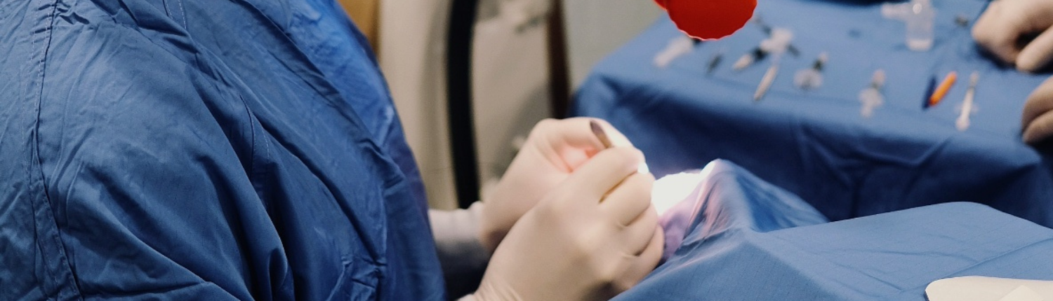
No. You’ll receive numbing eye drops and mild sedation. Most patients experience no pain and little to no memory of the procedure. Slight irritation or scratchiness may occur afterward but typically resolves quickly.
How long does the surgery take?
Each eye takes about 20 minutes. You’ll rest afterward and be discharged the same day.
Will I be awake during my cataract surgery?
Yes, but you’ll be comfortable and relaxed. Most patients feel no discomfort and are surprised by how quick and easy the experience is.
Before & After Surgery
Can I have cataract surgery the same day as my consultation?
No. Proper planning is essential. Custom lenses need to be ordered after advanced biometric measurements are taken. Once payment is made, your lenses are ordered and your surgery date is confirmed.
Can both eyes be done on the same day?
Only under special conditions, it is often better to allow one eye to recover before having the second eye done.
Do I need someone to drive me home?
Yes. You must arrange transport, as you won’t be able to drive immediately after surgery.
Recovery & Healing
What is the recovery timeline after cataract surgery?
- Vision: Vision often improves within 1–2 days, with full clarity over the following weeks.
- Screen Use: Light screen work is fine after 1–2 days, but take breaks and use lubricating drops.
- Exercise: Walking is allowed after a few days. Avoid intense workouts for 2 weeks.
- Swimming: No swimming or hot tubs for 4 weeks.
- Face Washing: Avoid washing your face on day 1. After that, use a clean cloth and warm (boiled) water. Pat dry—don’t rub.
- Hair Washing: You can resume after a few days. Tilt your head back to avoid water in your eyes.
- Makeup & Perfume: No makeup or perfume on the day of surgery or the first 24 hours. Avoid eye makeup for 2 weeks.
What are the risks or complications of cataract surgery?
Cataract surgery is safe and effective. Rare complications include infection, inflammation, retinal detachment, or swelling. You’ll be monitored closely post-op to catch any issues early.
Costs & Medical Aid
Is cataract surgery covered by medical aid?
Yes, it’s a Prescribed Minimum Benefit (PMB). However, medical aids usually only cover basic monofocal lenses. Premium multifocal or toric lenses may involve a co-payment. We’ll provide clear quotes and help with authorization.
Will I have out-of-pocket costs?
Possibly, if you opt for advanced lens technology or if your plan doesn’t cover all diagnostic tests. Our team will guide you through financial options and assist with reimbursement documents.
Surgical Retinal Conditions:

Understanding Surgical Retinal Conditions
What are surgical retinal conditions?
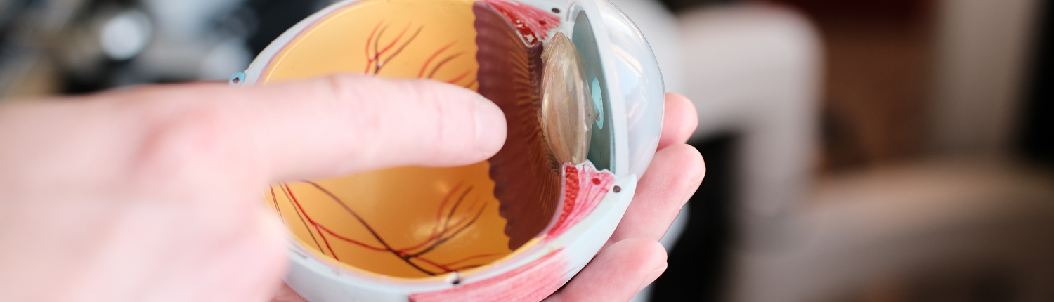
Surgical retinal conditions affect the retina, the light-sensitive layer of tissue at the back of the eye, and often require delicate surgical intervention to save or improve vision. They may be caused by aging, trauma, diabetes, or inherited diseases.
Which retinal conditions does Dr. Dean treat surgically?
Dr. Dean van der Westhuizen is skilled in a wide range of advanced retinal procedures, including:
- Retinal detachment repairs (primary, complex, or traumatic)
- Diabetic vitrectomy for proliferative diabetic retinopathy
- Macular hole surgery
- Epiretinal membrane (ERM) peeling
- Vitreomacular traction release
- Intraocular foreign body removal (including via modified lasso technique)
Other conditions managed include:
- Age-related macular degeneration (AMD)
- Diabetic macular oedema (DME)
- Central serous chorioretinopathy (CSCR)
- Choroidal neovascular membranes (CNVM)
- Inherited or dystrophic retinal diseases
- Inflammatory retinal disorder
What symptoms might indicate a retinal problem?
- Sudden increase in floaters (tiny specks in vision)
- Flashes of light in peripheral vision
- A shadow or curtain effect in your field of vision
- Distorted or blurry central vision
- Sudden or progressive loss of vision
These may be signs of serious retinal problems such as retinal detachment or macular disease. Prompt diagnosis is essential.
Book an Urgent Retina Evaluation →
How are retinal conditions diagnosed?
Diagnosing retinal disease involves specialized imaging and examination techniques, including:
- OCT (Optical Coherence Tomography): Cross-sectional scans of the retina
- Fundus Photography: High-resolution images of the retina and optic nerve
- Fluorescein Angiography: Evaluates blood flow in retinal vessels
- Ultrasound B-scan: Used when internal structures are obscured by hemorrhage or media opacity
- Widefield Imaging: Captures peripheral retina for a full diagnostic picture
Early detection can prevent irreversible vision loss. Schedule a Diagnostic Assessment →
Surgical Retinal Treatments
How are surgical retinal conditions treated?
Treatment depends on the condition and severity. Surgical options include:
- Vitrectomy: Removal of the vitreous gel to access the retina and relieve traction
- Laser Photocoagulation: Seals retinal tears or bleeding vessels
- Scleral Buckling: External support for detached retina
- Membrane Peeling: Removes scar tissue on the macula
- Gas or oil tamponade: Used to support the retina post-surgery
How long does retinal surgery take?
Most retinal surgeries take 20–60 minutes per eye, depending on complexity.
Will I be awake during the retinal procedure?
Yes, but you will receive sedation and numbing medication. Most patients are relaxed and pain-free during surgery.
Can I have surgery on the same day as my consultation?
No. Surgery requires advanced planning, diagnostic imaging, and custom surgical strategy. Once measurements are complete, a surgery date will be scheduled.
Can both eyes be operated on the same day?
In some cases, yes. But often, for safety and clarity of healing, surgeries are staged. We’ll help navigate medical aid protocols and recovery planning.
Post-Surgery & Recovery
What is the recovery like after retinal surgery?
Recovery depends on the procedure and whether a gas/oil bubble is used. General guidelines:
What You Can Expect:
- Vision: Blurry vision initially. It improves gradually over days to weeks.
- Discomfort: Mild scratchiness or pressure is common. Severe pain is not.
- Positioning: You may need to posture face-down (especially with gas tamponade).
- Follow-up: Your eye will be monitored closely in the weeks that follow.
Activity Guidelines:
- Screen time: Limited screen use is fine within 1–2 days, but take breaks.
- Exercise: Light walking may resume within a few days. No heavy lifting, yoga, or contact sports for 2–4 weeks.
- Swimming: Avoid for 4–6 weeks.
- Flying: You must not fly if you have a gas bubble in your eye.
- Face washing: Avoid splashing for 1–2 days. Clean gently with boiled lukewarm water and a cotton pad.
- Makeup: No makeup for 1 week; no eye makeup for at least 2 weeks.
- Hair washing: Resume with care, keeping water and shampoo out of your eyes.
You’ll receive personalized recovery instructions depending on the procedure.
Download Our Retina Surgery Aftercare Guide →
Financial Questions
Will medical aid cover my retinal surgery?
Yes, most retinal surgeries are covered under Prescribed Minimum Benefits (PMBs). However:
- Diagnostic scans may be billed from your day-to-day or hospital benefit
- Premium consumables (e.g., certain tamponade agents or instruments) may require co-payments
- Our practice will assist you with quotations, motivation letters, and authorizations
Can I get a quote before surgery?
Absolutely. We provide transparent, detailed quotes and can assist with prior approval where required.
You’re not just a case—you’re a person with a vision to protect.
Meet Dr. Dean →
Medical Retinal Conditions:


Understanding Medical Retinal Conditions
What are medical retinal conditions?
Medical retinal conditions are diseases that affect the retina—the light-sensitive tissue at the back of the eye—but are typically treated with medications, intravitreal injections, laser therapies, or observation rather than surgery. These conditions often arise from aging, systemic diseases like diabetes or hypertension, inflammation, or genetic predisposition. Some may eventually require surgery, but most are managed using specialized medical protocols and close follow-up.
What specific medical retinal conditions are treated at your practice?
Our team manages a comprehensive spectrum of medical retinal diseases, including:
- Age-related Macular Degeneration (AMD)
– Dry (atrophic) and wet (neovascular) forms
– Intravitreal anti-VEGF therapy for wet AMD - Diabetic Retinopathy & Diabetic Macular Oedema (DME)
– Screening, monitoring, and treatment with laser or injections
– Pan-retinal photocoagulation (PRP) and intravitreal therapy - Central Serous Chorioretinopathy (CSCR)
– Observation, medical treatment, or subthreshold laser when needed - Choroidal Neovascular Membranes (CNVM)
– Often secondary to AMD, high myopia, or inflammation
– Managed with intravitreal injections and imaging follow-up - Retinal Vein Occlusions (BRVO, CRVO)
– Macular oedema and neovascular complications are treated with anti-VEGF or steroid injections - Retinal Dystrophies & Inherited Retinal Disorders
– Including retinitis pigmentosa, cone-rod dystrophies, and Stargardt disease
– Managed with genetic counselling, visual rehabilitation, and low-vision support - Inflammatory and Autoimmune Retinopathies
– Includes posterior uveitis, birdshot chorioretinopathy, and sarcoidosis-related conditions
– Requires systemic evaluation and immunosuppressive therapy in collaboration with rheumatology - Macular Telangiectasia (MacTel)
– Monitored with OCT and managed when complications like CNVM arise - Floaters and Posterior Vitreous Detachment (PVD)
– Assessed for underlying retinal tears or detachment
If you’ve been diagnosed with any of these conditions—or are experiencing unexplained vision changes—we encourage a prompt and thorough assessment.
Book a Medical Retina Consultation →
What symptoms might suggest a medical retinal condition?
- Blurred or distorted central vision
- Wavy lines (metamorphopsia) when looking at straight edges
- Dull or faded colors
- New floaters (small moving specks or shadows in your vision)
- Flashes of light (especially in the periphery)
- A sudden loss of vision or a dark “curtain” in your field of view
- Difficulty reading or recognizing faces
Sudden onset symptoms, particularly floaters and flashes, may indicate retinal tears or detachment and require urgent attention.
Call Immediately for Urgent Triage →
How are medical retinal conditions diagnosed?
We use state-of-the-art diagnostic tools to detect, monitor, and manage retinal diseases. These include:
- Optical Coherence Tomography (OCT): High-resolution cross-sectional imaging of the retina
- OCT Angiography (OCTA): Visualizes blood vessels in the retina without dye
- Fluorescein Angiography (FFA): Dye-based imaging to identify leaking vessels or neovascularization
- Indocyanine Green Angiography (ICG): Used to evaluate deeper layers of the choroid
- Widefield Fundus Photography: Captures panoramic views of the retina
- Ultrasound B-scan: Useful in dense cataract or hemorrhage
- Visual Field Testing (VF): Evaluates functional defects in central and peripheral vision
- Fundus Autofluorescence: Assesses retinal pigment and metabolic stress
- Electroretinography (ERG) and Visual Evoked Potential (VEP): Functional testing for inherited or neurological disorders
- Corneal Topography & Wavefront Analysis: Used to rule out refractive or anterior segment causes when evaluating visual complaints
How are medical retinal conditions treated?
Treatment varies depending on the condition, severity, and cause. Options include:
- Intravitreal Injections (Anti-VEGF or Steroids):
– Used for wet AMD, DME, vein occlusions, and CNVM
– Delivered in our sterile, in-office injection suite - Laser Therapy:
– Pan-retinal photocoagulation for proliferative diabetic retinopathy
– Grid or focal laser for macular oedema
– Subthreshold laser for CSCR and macular leaks - Oral or Topical Medication:
– Used in inflammatory and vascular conditions - Systemic Collaboration:
– We work with endocrinologists, rheumatologists, and neurologists when systemic diseases are involved - Observation and Monitoring:
– Many conditions like dry AMD or mild CSCR are best managed with regular imaging and lifestyle adjustments
How often will I need to come for treatment?
Treatment schedules vary. Some patients may need monthly visits for injections, while others are seen every few months for monitoring. Our goal is to use the fewest treatments necessary to stabilize and preserve your vision.
Diagnosis, Investigations & Treatment Options
How are medical retinal conditions diagnosed?
We use state-of-the-art diagnostic tools to detect, monitor, and manage retinal diseases. These include:
- Optical Coherence Tomography (OCT): High-resolution cross-sectional imaging of the retina
- OCT Angiography (OCTA): Visualizes blood vessels in the retina without dye
- Fluorescein Angiography (FFA): Dye-based imaging to identify leaking vessels or neovascularization
- Indocyanine Green Angiography (ICG): Used to evaluate deeper layers of the choroid
- Widefield Fundus Photography: Captures panoramic views of the retina
- Ultrasound B-scan: Useful in dense cataract or hemorrhage
- Visual Field Testing (VF): Evaluates functional defects in central and peripheral vision
- Fundus Autofluorescence: Assesses retinal pigment and metabolic stress
- Electroretinography (ERG) and Visual Evoked Potential (VEP): Functional testing for inherited or neurological disorders
- Corneal Topography & Wavefront Analysis: Used to rule out refractive or anterior segment causes when evaluating visual complaints
How are medical retinal conditions treated?
Treatment varies depending on the condition, severity, and cause. Options include:
- Intravitreal Injections (Anti-VEGF or Steroids):
– Used for wet AMD, DME, vein occlusions, and CNVM
– Delivered in our sterile, in-office injection suite - Laser Therapy:
– Pan-retinal photocoagulation for proliferative diabetic retinopathy
– Grid or focal laser for macular oedema
– Subthreshold laser for CSCR and macular leaks - Oral or Topical Medication:
– Used in inflammatory and vascular conditions - Systemic Collaboration:
– We work with endocrinologists, rheumatologists, and neurologists when systemic diseases are involved - Observation and Monitoring:
– Many conditions like dry AMD or mild CSCR are best managed with regular imaging and lifestyle adjustments
How often will I need to come for treatment?
Treatment schedules vary. Some patients may need monthly visits for injections, while others are seen every few months for monitoring. Our goal is to use the fewest treatments necessary to stabilize and preserve your vision.
Glaucoma:

Understanding Glaucoma
What is glaucoma?
Glaucoma is a group of eye diseases that damage the optic nerve—the vital connection between your eye and brain. This damage is often caused by elevated intraocular pressure (IOP) but may also occur even with normal pressure levels. Without treatment, glaucoma can lead to permanent vision loss, typically starting in the peripheral (side) vision.
Is glaucoma curable?
No. Glaucoma cannot be cured, but it can be managed. With early diagnosis and lifelong treatment, further damage to your vision can often be prevented.
Who is at risk of developing glaucoma?
You are at higher risk if you:
- Are over 40 years old
- Have a family history of glaucoma
- Are diabetic or have high blood pressure
- Are very nearsighted (myopic)
- Have had eye trauma or use long-term corticosteroids
- Are of African descent (higher risk of certain types)
If you’re over 40, especially with a family history, regular screening is essential.
Book a Glaucoma Screening →
What are the main types of glaucoma?
- Primary Open-Angle Glaucoma (POAG):
The most common form. It develops slowly and painlessly, often without symptoms until significant vision is lost. - Angle-Closure Glaucoma:
A sudden rise in eye pressure due to blocked drainage angle. It’s a medical emergency that can cause rapid vision loss and severe eye pain. - Normal-Tension Glaucoma:
Optic nerve damage despite “normal” eye pressure. Believed to be related to poor blood flow to the nerve. - Secondary Glaucoma:
Caused by other eye diseases, trauma, medications, or inflammation. - Congenital or Childhood Glaucoma:
Rare and typically diagnosed in infants or children. Requires surgical treatment.
How is glaucoma diagnosed?
At your glaucoma assessment, we will perform several key tests:
- Intraocular Pressure Measurement (Tonometry)
- Optic Nerve Exam (Ophthalmoscopy or OCT imaging)
- Visual Field Testing (Perimetry)
- Gonioscopy (Angle Evaluation)
- Pachymetry (Corneal Thickness Measurement)
- OCT Imaging (Optical Coherence Tomography) to measure the health of the optic nerve and retinal nerve fiber layer
We use advanced OCT and visual field tools for early detection and tracking progression.
How is glaucoma treated?
Glaucoma treatment focuses on lowering intraocular pressure to prevent further optic nerve damage. Options include:
1. Eye Drops
- First-line treatment
- Reduce fluid production or increase fluid outflow
- Require daily use and consistent follow-up
2. Laser Treatments
- SLT (Selective Laser Trabeculoplasty): Non-invasive, used for open-angle glaucoma
- Laser Iridotomy: Used for angle-closure glaucoma
- Often reduce the need for drops
3. Surgery
- Trabeculectomy or Glaucoma Drainage Devices (e.g. Ahmed Valve): For advanced or resistant cases
- Minimally Invasive Glaucoma Surgery (MIGS): Safer with faster recovery; increasingly used alongside cataract surgery
4. Combination with Cataract Surgery
Patients with both glaucoma and cataracts may benefit from combined procedures.
Can I drive if I have glaucoma?
Yes. Many people with glaucoma drive safely, especially in early stages. However, advanced glaucoma can affect peripheral vision. You’ll need regular assessments to determine fitness to drive.
Do I need to change my lifestyle?
Generally, you can live a normal life, but:
- Avoid medications that increase eye pressure unless advised
- Exercise regularly, but avoid inverted yoga poses or heavy lifting
- Keep medical appointments and take medications as prescribed
- Tell all your doctors you have glaucoma, especially if prescribed steroids
Can I go blind from glaucoma?
If untreated, yes. But if diagnosed early and treated consistently, vision can often be preserved for life.
Financial Questions
Will my medical aid cover glaucoma treatment?
Yes, most treatments, drops, laser, and surgery are covered under Prescribed Minimum Benefits (PMBs). Some medications or surgical devices may require co-payments depending on your scheme.
Can I get a quote for surgery or laser?
Absolutely. Our admin team will provide detailed quotes and assist with pre-authorisation where needed.
Our goal is not just to manage your pressure, it’s to protect your quality of life.
Pterygiums:

Understanding Pterygiums
What is a pterygium?
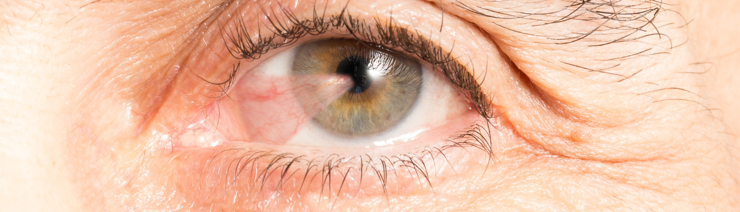
A pterygium is a benign (non-cancerous) growth of fibrovascular tissue on the surface of the eye. It typically starts on the white of the eye (conjunctiva) and may extend toward or across the cornea (the clear front part of the eye). It often appears triangular or wing-shaped.
Is a pterygium the same as a “surfer’s eye”?
Yes, “surfer’s eye” is a nickname for pterygium. It’s commonly seen in people who spend a lot of time outdoors in the sun, wind, and dust—especially near reflective surfaces like water or sand.
What causes pterygiums?
- Chronic UV light exposure
- Wind, dust, or environmental irritation
- Dry eye and inflammation
- Genetics (some people are more prone)
Who is at risk?
- People who work or play outdoors (farmers, surfers, cyclists, hikers)
- Those living in sunny or dry climates
- Patients with a family history of pterygium
- Individuals with dry or allergy-prone eyes
What are the symptoms of a pterygium?
- Red, inflamed or itchy eyes
- Foreign body sensation or irritation
- Grittiness or dryness
- Cosmetically visible growth on the white of the eye
- Blurred or distorted vision (if it encroaches onto the cornea)
Can a pterygium affect vision?
Yes. If the growth reaches the central cornea or induces astigmatism by pulling on the corneal surface, it can blur or distort your vision. Rarely, large or recurrent pterygiums can scar the cornea.
How do I know if it’s a pterygium or something more serious?
Pterygiums are usually diagnosed during a routine eye exam. In some cases, we may perform imaging or take photographs to document the growth. It’s important to rule out other conditions like pinguecula, conjunctival tumors, or scleral thinning.
Do all pterygiums require surgery?
No. If the pterygium is small, not growing, and not causing symptoms, it can be monitored without surgery.
How are pterygiums managed without surgery?
- Lubricating drops to reduce dryness and irritation
- Anti-inflammatory eye drops to reduce redness and swelling
- Sunglasses and UV protection to slow progression
- Avoidance of wind, dust, and allergens
- Allergy treatment if associated with itchy, watery eyes
Surgical Treatment
When is pterygium surgery recommended?
Surgery may be recommended if:
- The pterygium is growing toward the central cornea
- It is affecting or threatening your vision
- You have persistent discomfort or irritation
- You are unhappy with its cosmetic appearance
- Contact lens wear is difficult due to the pterygium
What does the surgery involve?
Pterygium surgery involves excising the abnormal tissue and covering the area with a conjunctival graft (often from the upper part of your own eye). This technique significantly reduces recurrence.
We use modern techniques, including:
- Sutureless surgery with tissue glue for faster healing
- Microsurgical tools for precision and comfort
- Conjunctival autograft or amniotic membrane as needed
Is the surgery painful?
No. The procedure is performed under local anesthesia, sometimes with light sedation. Mild irritation or scratchiness is common for a few days afterward, but pain is usually minimal.
What is recovery like?
- Mild redness, irritation, or light sensitivity is common in the first week
- Vision may be blurred for a few days
- You’ll use eye drops for 2–4 weeks to promote healing and prevent inflammation
- Most patients return to work within 3–5 days, depending on the environment
- Protective sunglasses are essential after surgery to prevent recurrence
Will the pterygium come back after surgery?
There is always a risk of recurrence, especially in young patients or those with heavy sun exposure. However, with modern surgical techniques like conjunctival autografting, recurrence rates are typically low (under 10%). Continued UV protection and proper eye care reduce your long-term risk.
Why Trust Us With Your Pterygium Care?
- Expert diagnosis and monitoring using high-resolution imaging
- Access to modern sutureless surgical techniques
- Personalized treatment plans balancing comfort, cosmesis, and safety
- Low recurrence rates and high patient satisfaction
- Supportive post-op care and education
- Professional guidance to protect your eyes long-term
Cost and Medical Aid
Is pterygium surgery covered by medical aid?
Yes, pterygium surgery is usually covered under most medical aid plans. Our team will provide a detailed quote and help with authorisation if needed. Cosmetic removal without medical indication may have out-of-pocket costs.
Can I get a cost estimate before booking surgery?
Absolutely. We’ll provide a transparent breakdown including consultation, surgical fees, and theatre charges.
Corneal Conditions:

Dry Eye Disease (DED)
What is dry eye disease?
Dry eye disease occurs when your eyes don’t produce enough tears, or when the tears evaporate too quickly. This leads to inflammation and discomfort, and can impair vision if left untreated.
What causes dry eyes?
- Age-related tear decline
- Hormonal changes (especially in women)
- Long hours of screen time or reading
- Air conditioning, wind, or dry climate
- Contact lens use
- Certain medications (e.g., antihistamines, antidepressants)
- Autoimmune diseases (e.g., Sjögren’s syndrome)
What are the symptoms of dry eyes?
- Gritty, sandy, or burning sensation
- Eye fatigue
- Intermittent blurry vision that improves with blinking
- Watery eyes (a paradoxical reaction)
- Light sensitivity
- Redness or irritation
How is dry eye diagnosed?
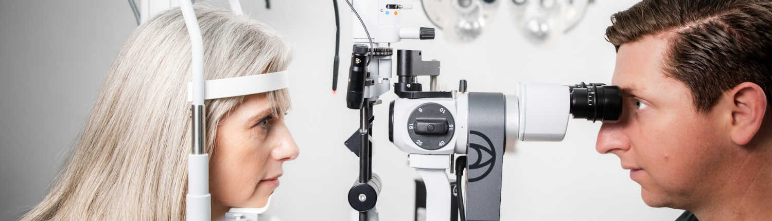
We assess your tear film using slit-lamp examination, fluorescein staining, and tear breakup time (TBUT). If needed, Schirmer’s test is used to measure tear production.
What are the treatment options for dry eyes?
- Preservative-free artificial tears
- Warm compresses and lid hygiene
- Anti-inflammatory drops (e.g. ciclosporin or corticosteroids)
- Punctal plugs to reduce tear drainage
- Omega-3 supplementation
- Lifestyle changes (e.g., reducing screen time, humidifiers)
- In-office treatments like thermal pulsation for meibomian gland dysfunction
Persistent dry eye affects both comfort and quality of vision. Don’t ignore it.
Book a Dry Eye Assessment →
Ocular Allergy
What is ocular allergy?
Ocular allergy (allergic conjunctivitis) is an immune response in the eye, often caused by environmental allergens like dust, pollen, mold, or animal dander.
What are the symptoms?
- Itchy, red, watery eyes
- Swollen eyelids
- Burning or foreign body sensation
- Stringy mucous discharge
- Symptoms often worsen during spring or windy seasons
How is it treated?
- Avoiding triggers (e.g., pollen exposure)
- Cold compresses
- Topical antihistamines and mast cell stabilizers
- Lubricating drops to wash out allergens
- Short-term corticosteroid drops (for severe cases)
- Oral antihistamines (with caution, as they may worsen dry eye)
Ocular allergy can mimic or worsen dry eye—accurate diagnosis is key.
Book an Allergy Eye Consultation →
Keratoconus
What is keratoconus?
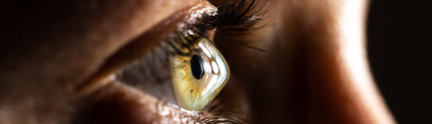
Keratoconus is a progressive eye condition where the cornea becomes thin and bulges outward into a cone shape. This distorts vision and may lead to scarring if untreated.
What causes it?
- Genetic predisposition
- Eye rubbing (often related to allergy or habit)
- Underlying collagen disorders
- Often begins in teens or early adulthood
What are the symptoms?
- Blurred, distorted, or ghosted vision
- Frequent prescription changes (especially increasing astigmatism)
- Sensitivity to light
- Difficulty with night vision
- Poor response to glasses
How is keratoconus diagnosed?
Diagnosis is made via corneal topography, which maps the shape and curvature of the cornea. Pentacam or Orbscan imaging helps detect early or subclinical keratoconus.
How is keratoconus treated?
- Early Stage: Spectacles or soft contact lenses
- Moderate Stage: Rigid gas-permeable or scleral contact lenses
- Progressive Stage: Corneal cross-linking (CXL) to halt progression
- Advanced Stage: Corneal transplant (deep anterior lamellar keratoplasty or full-thickness graft)
Early detection and cross-linking can preserve vision and delay or avoid surgery.
Book a Keratoconus Screening →
Corneal Cross-Linking (CXL)
What is corneal cross-linking?
Cross-linking is a minimally invasive treatment that strengthens the cornea to stop the progression of keratoconus and related ectasias.
How does it work?
A combination of ultraviolet (UV) light and riboflavin (vitamin B2) is used to create new collagen bonds in the cornea, making it more rigid and stable.
Is cross-linking painful?
Mild discomfort is common for 3–5 days after the procedure. You’ll wear a bandage contact lens and receive pain-relieving and antibiotic drops.
Does cross-linking improve vision?
Cross-linking is not primarily aimed at improving vision—it’s designed to stop worsening. However, in some cases, vision may improve slightly over time
When should cross-linking be done?
As soon as progression is detected—especially in young patients. Delayed treatment may lead to scarring or the need for a corneal transplant.
Cross-linking can preserve lifelong visual function if done early.
Schedule a Cross-Linking Consultation →
Corneal Transplantation
When is a corneal transplant needed?
Corneal transplant (keratoplasty) may be required if the cornea becomes severely scarred, distorted, or swollen, impairing vision. Indications include:
- Advanced keratoconus
- Corneal scarring (infection, trauma, or chemical injury)
- Fuchs’ endothelial dystrophy
- Failed previous grafts
- Corneal ulcers or perforation
What types of transplants are available?
- Penetrating Keratoplasty (PK): Full-thickness corneal transplant
- DALK (Deep Anterior Lamellar Keratoplasty): Replaces front layers, preserving the inner cornea
- DSAEK/DMEK: For endothelial failure—replaces only the damaged inner layer
What is the recovery time?
- Vision improves gradually over months
- Sutures may remain for up to a year
- Regular follow-up is essential to monitor healing and graft health
- Anti-rejection eye drops are used long-term
What is the success rate?
Corneal transplants are highly successful. Success depends on the indication, graft type, and patient compliance.
If your cornea has become too damaged or irregular for lenses or cross-linking, surgery may restore clarity.
Book a Corneal Surgery Consultation →
Uveitis:

Understanding Uveitis
What is uveitis?
Uveitis is inflammation of the uvea—the middle layer of the eye that includes the iris, ciliary body, and choroid. However, the term is also used broadly to describe inflammation inside the eye that may affect the retina, vitreous, or optic nerve.
What are the types of uveitis?
Uveitis is categorized by where the inflammation occurs:
- Anterior Uveitis (Iritis): Affects the front of the eye (iris); most common and often painful
- Intermediate Uveitis: Affects the vitreous (gel-like fluid in the eye)
- Posterior Uveitis: Affects the retina or choroid at the back of the eye
Is uveitis serious?
Yes. Uveitis can lead to permanent vision loss if not treated early and effectively. It may also cause complications such as glaucoma, cataracts, macular oedema, and retinal damage.
What are the symptoms of uveitis?
Symptoms vary based on the type, but may include:
- Red, painful eye
- Blurred vision
- Light sensitivity (photophobia)
- Floaters (dark spots in vision)
- Eye tenderness or watering
- Decreased vision, especially in posterior uveitis
If you have any of these symptoms, seek an urgent evaluation—early treatment protects sight.
Book an Urgent Eye Exam →
What causes uveitis?
In many cases, no specific cause is found (idiopathic). However, possible causes include:
- Autoimmune diseases:
– Rheumatoid arthritis
– Ankylosing spondylitis
– Sarcoidosis
– Lupus
– Inflammatory bowel disease (Crohn’s, ulcerative colitis) - Infections:
– Tuberculosis
– Toxoplasmosis
– Herpes simplex or zoster
– HIV, CMV, or syphilis - Post-surgical inflammation or trauma
- Genetic predisposition (e.g., HLA-B27 positive individuals)
Is uveitis linked to systemic health problems?
Yes. Up to 50% of cases may be associated with systemic inflammatory or autoimmune diseases. Identifying this link can guide long-term treatment and prevent future flares.
Treatments
How is uveitis diagnosed?
Diagnosis involves a thorough eye examination and imaging tests:
- Slit-lamp examination to detect anterior chamber cells and flare
- Fundoscopy to evaluate the retina and choroid
- Optical Coherence Tomography (OCT) for macular oedema
- B-scan ultrasound (if view is obscured)
- Fundus fluorescein angiography or ICG angiography for vascular leakage
Will I need blood tests or scans?
Yes, if systemic disease is suspected, you may need:
- Blood tests (e.g., ANA, HLA-B27, ACE levels, ESR, CRP)
- Chest X-ray or CT scan (for TB or sarcoidosis)
- Infectious disease screening
- Rheumatology or infectious disease referral
How is uveitis treated?
Treatment depends on severity and cause:
First-line therapies:
- Topical steroid drops (e.g., Prednisolone): For anterior uveitis
- Cycloplegic drops (e.g., Atropine): To reduce pain and prevent complications
For moderate/severe or posterior disease:
- Oral corticosteroids (Prednisone)
- Intravitreal steroid injections
- Immunosuppressive medications (e.g., methotrexate, azathioprine, biologics)
– Managed with input from a rheumatologist or physician - Antibiotics or antivirals (if infectious cause is found)
Is uveitis curable?
Acute uveitis can often be controlled completely. Chronic or recurrent uveitis may need long-term monitoring and treatment to prevent flare-ups and vision loss.
Will I need regular follow-up visits?
Yes. Uveitis can recur or worsen silently. Regular monitoring allows your ophthalmologist to:
- Adjust medications
- Detect complications early (glaucoma, cataract, macular oedema)
- Track visual recovery
Can uveitis cause permanent damage?
Without prompt and appropriate treatment, yes. Complications may include:
- Cataract formation
- Glaucoma
- Macular oedema
- Retinal scarring or detachment
- Vision loss
With proper care, many patients retain good vision and lead normal lives.
Emergency Eye Conditions & Ocular Trauma:

Eye Emergency
What qualifies as an eye emergency?
Any eye condition that threatens vision or eye integrity is considered an emergency. This includes:
- Sudden loss or decrease of vision
- Trauma or injury to the eye
- Chemical exposure
- Foreign bodies in the eye
- Eye pain with redness or light sensitivity
- Flashes and floaters with shadow or curtain effect
- Sudden double vision or drooping eyelid
- Bleeding inside the eye (hyphema)
- Corneal ulcers or severe infections
If you’re unsure whether it’s an emergency—treat it as one until proven otherwise.
Call Now for Urgent Advice →
What are the most common types of eye injuries?
1. Blunt Trauma (e.g., fist, ball, dashboard injury)
- May cause bruising, orbital fractures, bleeding inside the eye (hyphema), or retinal detachment
- Symptoms: pain, blurred vision, swelling, floaters
Do not rub the eye. Seek immediate care.
2. Penetrating Injuries (e.g., metal shards, glass, sharp objects)
- Serious and vision-threatening
- Do not attempt to remove objects yourself
- Shield the eye and go directly to emergency care
3. Chemical Exposure
- Alkaline agents (e.g., oven cleaner, cement) are especially dangerous
- Irrigate immediately with clean water or saline for 10–20 minutes
- Remove contact lenses
- Then seek immediate medical care
4. Foreign Bodies (e.g., dust, metal filings, wood splinters)
- May cause corneal abrasions or rust rings
- Symptoms: tearing, blinking, foreign body sensation
- Do not rub the eye
- Come in for removal and antibiotic protection
5. Thermal Burns
- Often from hot steam or metal
- Require urgent cooling, lubrication, and ophthalmic evaluation
What medical eye conditions need urgent treatment?
Retinal Detachment
- Sudden flashes, floaters, or curtain over vision
- Risk of permanent vision loss if untreated within 24–72 hours
Acute Angle-Closure Glaucoma
- Sudden pain, red eye, headache, nausea, blurred vision, halos
- Eye pressure spikes dangerously
- Requires immediate pressure-lowering treatment
Central Retinal Artery Occlusion (CRAO)
- Sudden, painless loss of vision in one eye
- “Eye stroke”—must be treated within hours
Endophthalmitis
- Severe infection inside the eye (often after surgery or injection)
- Redness, pain, vision loss, discharge
- Requires urgent intravitreal antibiotics
Corneal Ulcers
- Open sores on the cornea, often due to contact lens misuse
- Pain, tearing, blurred vision, white spot on the eye
- Needs prompt antibiotics to prevent scarring or perforation
Diagnosis & Treatments
What happens during an emergency eye exam?
Depending on your symptoms, we may perform:
- Slit-lamp examination
- Fluorescein staining (to detect corneal damage)
- Tonometry (eye pressure measurement)
- Retinal examination
- B-scan ultrasound (if the view is blocked)
- Anterior segment and fundus photography
- OCT and widefield imaging (if time and condition permit)
What emergency treatments are available?
- Eye irrigation (for chemical burns)
- Antibiotic or steroid eye drops
- Pressure-lowering medications for glaucoma
- Intravitreal injections (for infections or vascular events)
- Laser procedures (for retinal tears or angle-closure glaucoma)
- Suturing for lacerations
- Corneal glue or bandage lens for perforations
- Referral to theatre for urgent surgical repair (intraocular foreign body, rupture, etc.)
What should I do if I injure my eye or experience sudden symptoms?
- Do not rub or press on the eye
- Do not self-medicate with expired drops or ointments
- Shield the eye gently (especially in penetrating injuries)
- Flush with water if exposed to chemicals
- Remove contact lenses (unless embedded)
- Seek medical attention immediately
Can I go to a general emergency room?
If it’s after hours and vision is at risk, yes. But general ERs may not have ophthalmic tools or expertise. Contact our team first whenever possible.