
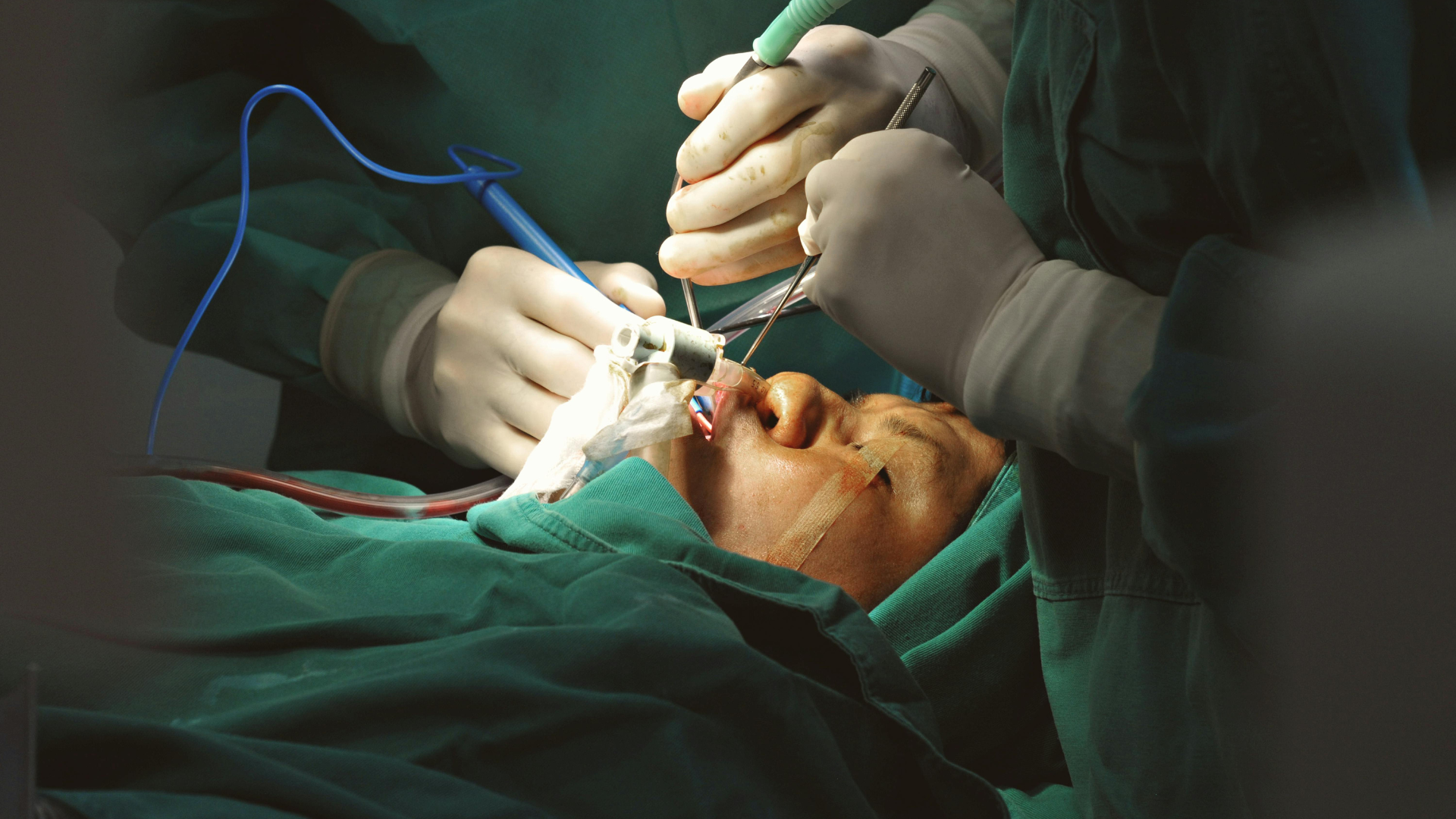
Retinal Detachment Surgery – What to Expect
A retinal detachment is one of the true emergencies in ophthalmology. If not treated timeously, it can lead to permanent vision loss. The good news is that with modern retinal surgery, many patients can have their sight preserved, especially if they are seen early.
As an eye specialist and eye surgeon at the Lowveld Eye Institute, I regularly manage retinal detachments for patients from across Mpumalanga. This article is based on my video “Retinal Detachment Surgery: What to Expect (Part 1)” and explains:
- Key symptoms you should never ignore
- Why retinal detachment happens
- The difference between a posterior vitreous detachment (PVD) and a retinal detachment
- How surgery works in practical terms
- Why timing matters so much for your final vision
What is a retinal detachment?
At the back of the eye lies the retina – a thin, delicate layer of nerve tissue that captures light and sends signals to the brain. You can think of it like the “film” or “sensor” in a camera.
A retinal detachment happens when:
- A tear or hole forms in the retina
- Fluid seeps through that break
- The retina lifts away from the underlying tissue and starts to peel off
Once the retina is detached, it cannot function properly. Without surgery, this can lead to severe and irreversible loss of vision.
Retinal detachment can happen to anyone, but it is more common in:
- Short-sighted (myopic) patients – especially with higher degrees of myopia
- Eyes that are longer than average, with abnormal adhesions between the vitreous gel and the retina
This is one of the reasons why regular checks with an eye specialist are so important if you are highly myopic.
Symptoms you should never ignore
Recognising early warning signs can make the difference between saving and losing central vision. Classic symptoms of a retinal tear or detachment include:
1. Flashes of light
- Often described as:
- “Lightning”
- “Sparklers”
- A brief crescent or arc of light in the periphery
- Typically:
- Worse at night
- Triggered by eye movements
- Visible even with eyes closed
Flashes suggest traction – the vitreous gel tugging on the retina.
2. New floaters
- Sudden appearance of:
- Dark spots
- Cobwebs
- A large ring or “blob” that moves with eye movement
- May be due to:
- The vitreous separating
- A small blood vessel tearing and bleeding into the gel
3. A dark curtain or shadow
This is the most worrying symptom:
- A black shadow or curtain that slowly spreads across your vision
- Can come from:
- Below
- Above
- One side
- Suggests that the retina is already detaching and the area of vision supplied by that part is switching off
If you experience flashes, new floaters or any curtain-like shadow, you should see an eye specialist or retinal surgeon as soon as possible. Waiting to “see if it goes away” can severely reduce your visual outcome.
Posterior vitreous detachment vs retinal detachment
It’s important not to confuse a posterior vitreous detachment (PVD) with a retinal detachment:
- Posterior vitreous detachment (PVD):
- The vitreous gel inside the eye naturally separates from the back of the eye with age
- Can cause flashes and floaters
- Is common and often benign, but can lead to a tear in some people
- Retinal detachment:
- Occurs when a tear forms and fluid tracks underneath the retina
- The retina lifts off, and vision is threatened
- Almost always requires surgery
If you’ve been told you have a PVD, this is not the same as a retinal detachment, but it does mean you should be monitored carefully, especially if you develop new symptoms.
How a retinal detachment develops
Inside the eye is the vitreous, a clear gel that fills the back chamber.
Over time:
- The vitreous becomes more liquid and mobile.
- It starts to peel away from the back of the eye; this is the PVD.
- As it pulls away, it can:
- Simply separate, causing flashes and floaters only, or
- Tear the retina at a point of strong adhesion
If a tear forms:
- Fluid from the vitreous cavity can pass through the tear
- This fluid accumulates under the retina, lifting it off like wallpaper peeling from a wall
- Detachment spreads over time, involving more and more of the retina
Once this process starts, surgery is usually the only way to reattach the retina and protect your vision.
Macula-on vs macula-off: why timing matters
One of the most important distinctions in retinal detachment is whether the macula, the central part of the retina responsible for fine, detailed vision, is still attached.
- Macula-on detachment
- The central retina is still attached
- Peripheral retina is detached
- If we operate before the macula detaches, the chances of maintaining good central vision are much higher
- Macula-off detachment
- The central retina has already lifted off
- Vision is usually significantly blurred or distorted
- Surgery is still essential, and vision can improve, but:
- Results are more variable
- Less predictable than when the macula is still on
This is why urgent assessment by an eye surgeon is so critical if you have warning symptoms. The earlier we intervene, the better the chance of preserving central vision.
How retinal detachment surgery works
Modern retinal detachment repair is most commonly done with a procedure called a pars plana vitrectomy (PPV). In simple terms, the aims are:
- Remove traction
- The vitreous gel that is tugging on the tear is removed (vitrectomy)
- Remove subretinal fluid
- The fluid under the detached retina is drained, allowing the retina to flatten back into place
- Seal the tear with laser
- Laser is applied around the retinal tear(s)
- Think of this like “spot welding”; it creates an adhesion so that the retina sticks down
- Support the retina from inside (tamponade)
- A gas or silicone oil bubble is placed in the eye
- This presses against the retina from the inside while the laser scars mature and become permanent
Gas vs oil tamponade: a brief overview
Your surgeon will decide between gas and oil based on:
- Type and complexity of the detachment
- Presence of scar tissue (PVR)
- Number and position of retinal tears
- How long the detachment has been present
- Your need to travel or fly in the coming weeks
Gas (air or special long-acting gases):
- Stays in the eye for days to weeks, then dissolves by itself
- No second surgery needed to remove it
- You cannot travel by air or change altitude significantly while gas is in the eye
- You will often see a moving line or “horizon” as the bubble shrinks and fluid fills in above it
Silicone oil:
- Does not dissolve by itself
- Usually requires a second surgery later to remove it
- You can fly and travel normally with oil in the eye
- Often used for more complex detachments or when stability is needed for a longer time
There is a separate dedicated explanation on gas vs silicone oil in retinal surgery, but this gives the basic reasoning behind the choice.
When to seek urgent help
You should urgently contact an eye specialist or eye doctor if you experience:
- New or worsening flashes of light
- Sudden onset of floaters, especially a shower of black spots
- A dark curtain, shadow or veil coming across your vision
- Sudden drop in vision in one eye
These symptoms don’t always mean you have a retinal detachment, but they are significant enough that they must be checked promptly.
Retinal detachment care in Nelspruit, Mbombela & Mpumalanga
Retinal detachment is a sight-threatening emergency, but with early diagnosis and modern surgery, many patients recover useful, and sometimes excellent, vision.
If you live in Nelspruit, Mbombela or the wider Mpumalanga region and are:
- Experiencing symptoms that worry you, or
- Have been told you have a retinal tear or detachment
You should seek assessment with an eye specialist/eye surgeon at the Lowfeld Eye Institute without delay.
Early action gives your retina and your vision the best possible chance.

